
Please note – this, like nearly all posts on this site, is generated by a large language model. While it may state many correct things, a healthy skepticism is warranted.
What Is Xenon MRI?
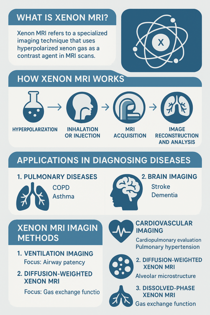
Xenon MRI refers to a specialized imaging technique that uses hyperpolarized xenon-129 gas as a contrast agent in MRI scans. Unlike conventional MRI, which focuses primarily on proton (hydrogen) signals, xenon MRI enables imaging of gas distribution and function, particularly in the lungs, brain, and other tissues where xenon dissolves and interacts with the environment.
Xenon-129 is a noble gas isotope that becomes hyperpolarized via spin-exchange optical pumping, enhancing its MRI signal by tens of thousands of times compared to its natural state. Once inhaled or introduced into the body, the xenon can be tracked to observe gas flow, uptake, and chemical interactions at a molecular level.
How Xenon MRI Works
- Hyperpolarization: Xenon gas is mixed with rubidium vapor and irradiated with laser light to align the nuclear spins of xenon atoms (boosting MRI signal).
- Inhalation or Injection: The gas is either inhaled (commonly for lung imaging) or administered in a controlled way for imaging other organs.
- MRI Acquisition: Specialized MRI sequences capture how xenon distributes in airspaces, diffuses into tissue, and interacts chemically with different compartments like blood plasma and red blood cells.
- Image Reconstruction and Analysis: The images show real-time functional information such as ventilation, gas exchange, and blood uptake.
Applications of Xenon MRI in Diagnosing Diseases
Xenon MRI is especially valuable in areas where traditional imaging techniques (like CT or standard MRI) offer limited functional or molecular information.
1. Pulmonary Diseases
Xenon MRI is most extensively used in lung imaging, due to xenon’s gaseous state and its ability to dissolve in lung tissue and blood.
- Chronic Obstructive Pulmonary Disease (COPD): Measures regional ventilation defects and gas transfer impairment.
- Asthma: Assesses ventilation heterogeneity and airway obstruction, even when spirometry is normal.
- Interstitial Lung Disease (ILD): Detects impaired gas exchange in early disease, differentiates from normal aging.
- Cystic Fibrosis: Reveals small airway disease and ventilation heterogeneity before structural damage appears.
- Post-COVID-19 Lung Dysfunction: Detects persistent impairment in gas exchange (even in normal CT scans).
2. Brain Imaging
Because xenon crosses the blood-brain barrier (BBB) and is lipophilic, it can be used to assess cerebral blood flow and gas exchange.
- Stroke & Ischemia: Measures regional cerebral perfusion non-invasively.
- Alzheimer’s & Dementia: Detects changes in BBB permeability and cerebral perfusion.
- Brain Tumors: Characterizes tumor vascularization and metabolism using xenon uptake and distribution.
3. Cardiovascular Imaging
- Cardiopulmonary Evaluation: Evaluates how well gas transfers from alveoli into blood, offering insight into heart-lung interaction.
- Pulmonary Hypertension: Provides a non-invasive window into gas exchange limitations due to vascular remodeling.
4. Other Emerging Applications
- Renal imaging (via dissolved-phase xenon): For evaluating perfusion and oxygenation.
- Oncology: Potential to detect tumor metabolism or drug delivery if xenon-tagged molecules are used.
Xenon MRI Imaging Methods
There are three main modes in which hyperpolarized xenon-129 is imaged:
1. Ventilation Imaging
- Focus: Airway patency and gas distribution.
- Method: Images inhaled xenon in lung airspaces.
- Application: COPD, asthma, CF.
2. Diffusion-Weighted Xenon MRI
- Focus: Alveolar microstructure.
- Method: Measures how xenon atoms diffuse through alveolar spaces.
- Application: Emphysema, ILD.
3. Dissolved-Phase Xenon MRI
- Focus: Gas exchange function.
- Method: Tracks xenon as it dissolves in tissues, blood plasma, and red blood cells.
- Application: ILD, pulmonary hypertension, brain perfusion.
Each compartment has a distinct chemical shift, allowing separation of xenon in airspaces (~0 ppm), tissue/plasma (~197 ppm), and RBCs (~217 ppm).
Criticisms and Limitations of Xenon MRI
1. Safety Concerns
- Inhalation Risks: While generally considered safe, high concentrations of xenon can be sedative and may depress respiratory drive. At diagnostic doses (~1/3 atmosphere), this is rarely problematic.
- Radiological Approval: Hyperpolarized xenon-129 is not yet FDA-approved for routine clinical use in many countries (as of 2025), limiting its adoption.
- Long-Term Effects: Repeated exposure risks are not fully characterized, though current evidence suggests low toxicity.
2. Technical Challenges
- Hyperpolarization Infrastructure: Requires specialized, expensive equipment (polarizers, gas handling systems).
- Short Signal Lifespan: The hyperpolarization decays quickly, requiring rapid transfer from preparation to imaging (~1-2 minutes).
- Low Signal-to-Noise Ratio (SNR): Despite hyperpolarization, xenon is present in much lower concentrations than hydrogen, making imaging more challenging.
- Complex MRI Sequences: Requires custom pulse sequences and expertise.
3. Cost and Accessibility
- High Cost: Due to xenon production, polarization, and custom imaging systems.
- Limited Availability: Found mostly in academic or research hospitals.
- Reimbursement Issues: Lack of insurance coding hampers widespread clinical use.
4. Clinical Utility Debates
- Critics argue that:
- Conventional tests (e.g., CT, spirometry, perfusion MRI) suffice in many cases.
- Xenon MRI often lacks outcome-based validation, i.e., does it meaningfully change treatment decisions?
- Lack of standardized protocols makes multicenter trials difficult.
Current Research and Future Directions
- Commercialization: Companies like Polarean Imaging are pushing for FDA approval of hyperpolarized xenon MRI, with promising trial results.
- Combination Imaging: Integrating xenon MRI with anatomical CT or proton MRI for comprehensive functional-structural insights.
- AI and Image Processing: Use of deep learning to improve image reconstruction and interpretation.
- Targeted Xenon Sensors: Use of xenon to detect pH, enzymatic activity, or receptor binding via cage molecules like cryptophanes — opening doors to molecular imaging and theranostics.
| Aspect | Details |
|---|---|
| Modality | Hyperpolarized xenon-129 MRI |
| Primary Use | Lung function imaging (ventilation, diffusion, gas exchange) |
| Other Uses | Brain perfusion, stroke, neurodegeneration, pulmonary hypertension |
| Strengths | Non-invasive, functional, regional imaging of gas transport and uptake |
| Limitations | High cost, limited access, regulatory hurdles, complex setup |
| Safety | Generally safe at clinical doses, mild anesthetic effects at high doses |
| Research Stage | Transitioning from research to clinical use in specialized centers |
Leave a Reply